
Clinical Significance of Ubiquitination Modification in the TAK1–TAB Complex
In the context of disease, the dysregulation of TAK1/TAB ubiquitination has been implicated in the pathogenesis of autoimmune conditions, tumor growth, and neurodegenerative diseases [8, 21, 61]. The excessive or prolonged activation of NF-
Ubiquitination, a pivotal post-translational modification, intricately regulates cellular processes critical for maintaining homeostasis [8, 64], including the cell cycle, proliferation, and apoptosis [65]. The ubiquitin–proteasome system (UPS) is crucial for protein degradation, similar to the regulatory roles of kinases [66, 67]. Disruptions in ubiquitination pathways can lead to an imbalance in these processes, significantly contributing to many disorders, including cancer, neurodegenerative diseases, and issues related to adaptive and innate immunity [68, 69]. Recent studies have underscored the dual role of ubiquitination in either promoting or inhibiting tumor progression, depending on the context and the specific proteins being targeted, while the UPS was also demonstrated as a potent target for anticancer therapy [67, 68, 70]. Deubiquitinating enzymes (DUBs) have emerged as critical regulators within this context, capable of removing ubiquitin modifications and, thus, reversing the effects on target proteins. Many DUBs have demonstrated tumorigenic roles in multiple cancers, and many inhibitors have been investigated in clinical trials for diverse cancer treatments [70]. Given their central role in modulating the stability and activity of key proteins involved in tumorigenesis, DUBs represent promising targets for therapeutic intervention, offering the potential for developing novel anticancer strategies [70]. However, the FDA has currently only approved a few drugs that target the ubiquitin system [66].
The advancements in ubiquitin biology have led to innovative strategies for modulating the ubiquitination system, with significant implications for therapeutic development [67]. For the past decade, proteasome inhibitors have been utilized in treating multiple myeloma (MM), yet resistance to these inhibitors has emerged, impacting their effectiveness. Bortezomib (Velcade) and carfilzomib (Kyprolis) are FDA-approved proteasome inhibitors effective against multiple myeloma [66]. Techniques such as proteolysis-targeting chimera (PROTAC) molecules [71] and hydrophobicity tags (HyT) [72] have been developed to specifically target and manipulate the degradation of proteins, offering precise approaches to treat diseases caused by dysregulation of the ubiquitination process [68]. Furthermore, the advent of small molecule inhibitors targeting ubiquitination enzymes presents a promising direction in treating conditions such as cancer, highlighting the potential of these molecular interventions in precision medicine [66].
Ubiquitination and phosphorylation are among the most crucial and widely studied post-translational modifications. It is a dynamic and versatile mechanism for controlling various aspects of cellular function [8, 9]. Ubiquitin, a small yet pivotal protein in cellular regulation, consists of 76 amino acids and is notable for its seven lysine residues—K6, K11, K27, K29, K33, K48, and K63—plus an N-terminal methionine (M1) that can all serve as attachment points for forming polyubiquitin chains [64, 73]. The functions and implications of ubiquitination through K6, K27, K29, and K33 are less well-characterized and represent an area ripe for further research [74, 75]. This ubiquitination system is a key regulator of intracellular proteins, influencing as much as 80% of them [70]. The consequences of ubiquitination can vary greatly (Fig. 4).
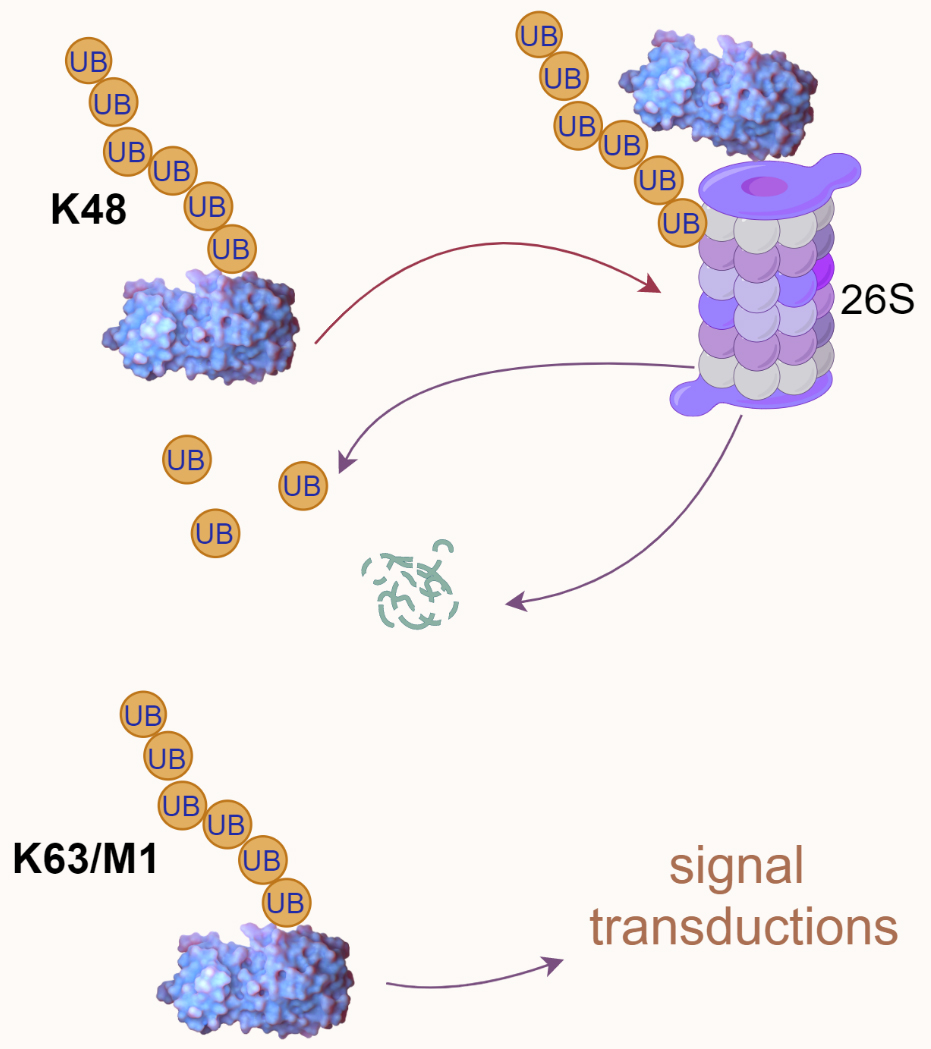 Fig. 4.
Ubiquitin-mediated proteasomal degradation and signal transduction. K48-linked polyubiquitination is typically associated with proteasomal degradation, as illustrated by a polyubiquitinated protein directed to the 26S proteasome. In contrast, K63/M1-linked polyubiquitination is often involved in signal transduction processes. UB, ubiquitin.
Proteasomal degradation: Ubiquitin chains, particularly those linked through the K48 residue, often signal the protein to be directed to the 26S proteasome for degradation. This is a key process for removing damaged, misfolded, or unneeded proteins, thereby regulating protein levels and quality control within the cell [73]. Altering substrate activity: Ubiquitination can also change the functional state of proteins, either activating or inhibiting their enzymatic activity, altering their cellular location, or affecting their ability to interact with other molecules [64]. Mediating protein–protein interactions: Ubiquitin chains can serve as a platform for assembling protein complexes, which is critical in various signaling pathways. For example, K63-linked ubiquitin chains are often involved in processes such as DNA repair, NF-
Ubiquitination encompasses non-proteolytic roles such as receptor internalization, multiprotein complex assembly, intracellular trafficking, and key signaling pathways, including those governing inflammation, autophagy, DNA repair, and enzymatic regulation [76, 77, 78]. Deregulation can trigger oncogenic pathways, disrupt cellular metabolism, and lead to inadequate protein complex formation critical for inflammation response or DNA repair, resulting in the accumulation of misfolded proteins, causing diseases such as neurodegeneration or misdirecting proteins away from their functional locations within the cell [8, 79].
Ubiquitination is a multistep process typically involving the coordinated action of three ubiquitination enzymes: ubiquitin-activating enzyme (E1), ubiquitin-conjugating enzyme (E2), and ubiquitin-protein ligase (E3) [64]. Initially, the E1 forms a high-energy thioester bond between the carboxyl group of the C-terminal lysine residue of ubiquitin and the thiol group of its own cysteine residue, utilizing ATP-provided energy. Subsequently, the activated ubiquitin is transferred onto the cysteine residue of the E2. Finally, a member of the highly conserved ubiquitin-protein ligase family, E3, recognizes specific target proteins to be ubiquitinated and catalyzes the transfer of ubiquitin molecules from E2 to the lysine residue of the target protein (Fig. 5) [64]. Really interesting new gene (RING) finger [RNF] proteins containing RING domains act as E3 ubiquitin ligases that mediate the covalent linkage of ubiquitin to target proteins [9]. Depending on the ubiquitin chain’s topology, ubiquitination can lead to proteasomal degradation or mediate protein–protein interactions, influencing various cellular processes [64]. Understanding these processes provides insights into the molecular mechanisms of diseases involving the TAK1–TAB complex and offers new opportunities for drug development.
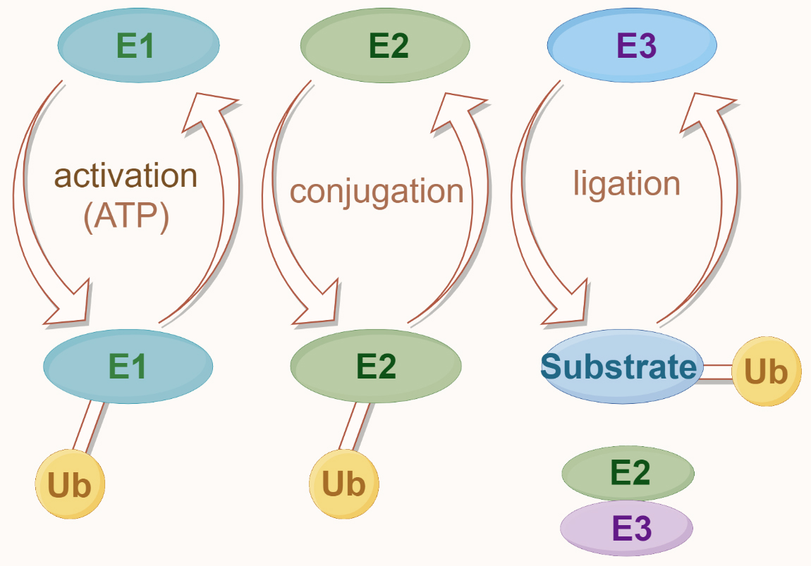 Fig. 5.
The ubiquitin modification system. Ubiquitination begins with the activation of ubiquitin (Ub) by the E1 enzyme, which is ATP-dependent. Activated ubiquitin is then transferred to the E2 conjugating enzyme. In the final step, the E3 ligase enzyme facilitates the attachment of ubiquitin to the substrate protein, completing the ubiquitination process.
TAK1 undergoes sophisticated regulation via different ubiquitin chain types, including K63-linked and K48-linked ubiquitination, each dictating unique biological outcomes [16]. E3 ubiquitin ligases, notably TRAF6 and TRAF2 [80, 81, 82, 83, 84], are pivotal in this regulation, intricately influencing TAK1’s cellular role. K63-linked polyubiquitination, identified at lysine residues K34, K158, K209, and K562, is crucial for TAK1 activation, with TRAF6 and TRAF2 mediation highlighting a complex regulatory network. A study by Fan et al. [81, 85] revealed that the activation of TAK1 via NF-
Recent investigations have expanded our understanding of TAK1 activation, revealing that its function may not solely depend on ubiquitination at known sites, suggesting a labyrinth of regulatory pathways yet to be explored. The tripartite motif-containing (TRIM) protein family [9], notably TRIM8 [86, 87] and TRIM27 [88], emerges as significant modulators within the TAK1/NF-
These investigations suggest that TAK1 activation is intricately modulated beyond known ubiquitination sites, hinting at a broader spectrum of regulatory pathways awaiting discovery. The involvement of the TRIM protein family, among other E3 ligases, in modulating TAK1 through diverse ubiquitination patterns underscores the multifaceted nature of its regulation. This complexity is crucial for maintaining cellular homeostasis and highlights the potential for targeted therapeutic interventions in diseases associated with TAK1 pathway dysregulation (Table 1, Ref. [57, 61, 80, 81, 82, 83, 84, 86, 87, 88, 89, 90, 91, 92, 93, 94, 95, 96, 97, 98, 99, 100, 101, 102, 103, 104]).
| Protein | Catalyzed by | Site | PTM | Reference |
| TAK1 | TRAF6 | K34, K158, K562 | K63 Ub | [80, 81, 82, 83, 84] |
| TRAF2 | K158 | K63 Ub | [82] | |
| TRIM8 | K158 | K63 Ub | [86, 87] | |
| Sef-S | K209 | K63 Ub | [89] | |
| CHIP | K63 Ub | [90] | ||
| ITCH | K72 | K48 Ub | [88, 92] | |
| TRIM31 | K72 | K48 Ub | [57] | |
| TRIM16 | K282, 547 | K48 Ub | [91] | |
| FBXW2 | K48 Ub | [93] | ||
| TRIM56 | M1 Ub | [94] | ||
| TAB1 | PHD | K294, K319, K335, K350 | K63 Ub | [61] |
| RNF207 | K63 Ub | [95] | ||
| ITCH | S452, S453, S456, S457 | K48 Ub | [96] | |
| TRIM26 | K294, K319 | K11 Ub | [97] | |
| K335 | ||||
| RNF114 | Ub | [98] | ||
| TAB2 | TRIM29 | K48 Ub | [99] | |
| RBCK1 | C673 | K48 Ub | [100] | |
| RNF99 | K611 | K48 Ub, | [101, 102] | |
| K63 Ub | ||||
| TRIM23 | Ub | [103] | ||
| TAB3 | AMFR | K649 | K27 Ub | [104] |
| RBCK1 | C692 | K48 Ub | [100] |
PTM, post-translational modification; K, lysine; C, cysteine; Ub, ubquitination.
TAB1, a critical regulator of TAK1 activity through direct interaction, is subject to a complex regulatory network via polyubiquitination and other post-translational modifications, including phosphorylation and O-GlcNAcylation [105, 106]. This multilayered modification landscape underscores the pivotal role of TAB1 in cellular signaling dynamics. Notably, both K63 and K48-linked ubiquitination have been observed on TAB1 [61, 95, 96], highlighting its versatile regulatory capabilities.
K63-linked ubiquitination of TAB1, particularly at lysine residues Lys294, Lys319, Lys335, and Lys350, is mediated by various E3 ligases, including the E3 ubiquitin ligase activity of the mitogen-activated protein kinase kinase 1 (MEKK1) the plant homeodomain (PHD) [61]. This modification is crucial for enhancing TAK1 aggregation and its kinase activity, significantly influencing embryonic stem cell differentiation and tumorigenesis. Intriguingly, recent discoveries, such as the work by Yuan et al. [95], have illuminated the role of RNF207 in activating the TAK1–JNK1/2 signaling pathway through K63-linked ubiquitination of TAB1, linking it to the exacerbation of stress-induced pathological cardiac hypertrophy.
TAB1 can also undergo K48-linked ubiquitination, although the specific modification sites remain unclear. Immune regulation is essential for maintaining skin integrity. Research by Theivanthiran et al. [96] suggests that ITCH can catalyze K48-linked polyubiquitination on TAB1, inhibiting the activation of p38
In addition to K63 and K48-linked ubiquitination, other types of ubiquitination also play crucial roles in TAB1’s functions. In a study by Zhao et al. [97], TRIM26 was found to catalyze K11-linked polyubiquitination on TAB1 at positions Lys294, Lys319, and Lys335, enhancing the activation of TAK1 and subsequent NF-
This enriched understanding of TAB1’s ubiquitination broadens our perspective on its regulatory functions and underscores the potential for targeting these modifications in therapeutic strategies, especially in contexts of cellular stress responses and disease pathogenesis.
Comments
Post a Comment